
Adie's syndrome consists of a tonic pupil and loss of deep tendon reflexes. Because the abnormality is post-ganglionic (that is, damage to the ciliary ganglion), one can demonstrate supersensitivity of the sphincter muscle to pilocarpine 1/10%.
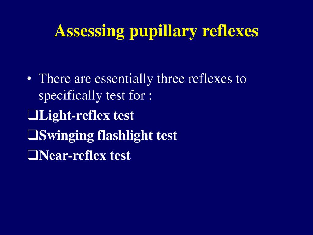
Under the slit lamp sectoral palsies of pupillary sphincter may be seen with light stimulation. Characterized by a dilated pupil with very poor or no light reaction with tonic constriction to near and tonic redilation. An isolated dilated pupil does not usually signify a III nerve palsy. A third nerve palsy will be usually associated with other signs of dysfunction including ocular motility disturbances and ptosis. Lesions of the parasympathetic system cause a dilated pupil Acute third nerve palsy May signal compression of the third nerve due to a posterior communicating artery aneurysm. In a normal eye the pupils should constrict Indirect light response Placing the penlight in front of the right eye (OD) The examiner watches the left eye for a consensual response.  The testing conditions for evaluating pupils include: Evaluate pupils in dim illumination Have the patient fixate at a distance (eliminates miosis from accomodation) Use bright light source Note size of pupils before assessing the responses look for anisocoria (unequal pupil size) and corectopia (irregular shaped pupil) Direct light response In watching the eye receiving the direct light, you can then determine the direct response by: Shining the penlight directly in the right, look at the response including speed of response note (brisk, sluggish) Repeating for the opposite eye If the pupils are normal, they will react equally to the direct light. With accommodative effort, a “near synkinesis” is evoked, including increased accommodation of the lens, convergence of the visual axes of the eyes and pupillary constriction or miosis. Changes of the pupil in accomodation: The pupil changes when focusing on near objects. Twenty percent of fibers in the optic tract are for pupillary function. Pupils are usually larger in children and smaller in the elderly. In dim lighting the pupil size is larger and in bright light it is smaller. The normal diameter of a pupil is 3 to 4 mm under average lighting conditions. Pupil Evaluation The pupil’s function is to control the amount of light entering the eyes, providing the best visual function under varying degrees of light intensity. Preventionĭue to the nature of the condition, there is no known cure or a way of preventing the disorder.Pupil assessment and abnormalities Pupillary light reflex pathway Relative afferent pupillary defect (RAPD) If medication is prescribed, the pet owner will need to ensure that all of the medication is given fully and as directed. Treatment will be fully dependent upon the underlying cause of the issue. Ultrasound can be used to detect lesions in the eyes, while computed tomography (CT) and magnetic resonance imaging (MRI) can be used to identify any lesions in the brain that may be causing the condition.
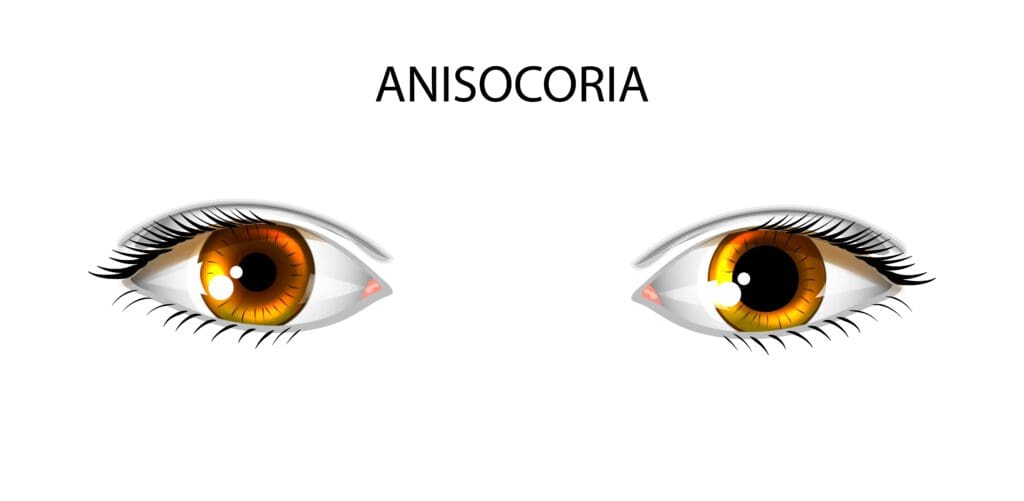
When veterinarians are evaluating the dog's pupils, the primary goal is to distinguish between neurological and eye-related causes. There are several potential causes of an altered pupil size in dogs, including inflammation in the frontal region of the eye, increased pressure in the eye, diseases that are focused in the iris tissue itself, a poorly developed iris, scar tissue build up in the eye, medications, and cancer. The most noticeable symptom is when your dog has one pupil that is visibly smaller than the other. If you would like to learn more about how this disease affects cats, please visit this page in the PetMD health library. The condition or disease described in this medical article can affect both dogs and cats. With the proper detection of the disease's underlying cause, treatment plans are available that should resolve the issue. This condition causes one of the dog's pupils to be smaller than the other. Anisocoria refers to an unequal pupil size. The pupil expands when there is little light present, and contracts when there is a greater amount of light present. The pupil is the circular opening in the center of the eye that allows light to pass through.


 0 kommentar(er)
